Transrectal ultrasound (TRUS) is regularly used to guide prostate biopsies, which constitute the current gold standard for cancer diagnosis. Unfortunately, TRUS suffers from low sensitivity, leading to a high rate of false negative results. Therefore, MR images – and more recently also Ga-labelled PSMA (Prostate Specific Membrane Antigen) PET – are used to identify suspicious areas in the prostate. The challenge is now to fuse pre-interventional PET/MRI with interventional TRUS both accurately enough to allow targeting with the biopsy needle and fast enough not to impede clinical routine with too much (time-consuming) user interaction.
In this project, we proposed a novel method to first automatically segment the prostate in TRUS using a Hough transform-based random forest approach (yes, this was before CNNs). Then, a elastic surface registration is performed to fuse (PET/)MRI with TRUS, relying on the Coherent Point Drift algorithm. The minimal computation time for the entire pipeline (less than five minutes) allows implementation in the clinical routine.
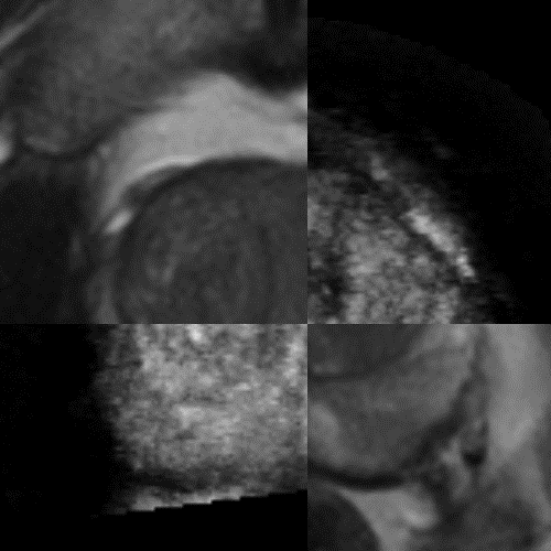
For biopsy guidance, the prostate is segmented in both TRUS and MRI images, and their surfaces are registered for a fusion visualization.
Related Publications
2017
Preconditioned intensity-based prostate registration using statistical deformation models
Oliver Zettinig, Julia Rackerseder, Beatrice Lentes, Tobias Maurer, Kay Westenfelder, Matthias Eiber, Benjamin Frisch, and Nassir Navab
In 2017 IEEE 14th International Symposium on Biomedical Imaging (ISBI 2017), Apr 2017
PET/MRI/TRUS image fusion guided prostate biopsy: Development of a research platform and initial clinical results
Benjamin Frisch, Enno Storz, Oliver Zettinig, Amit Shah, Hubert Kübler, Nassir Navab, Hans-Jürgen Wester, Markus Schwaiger, Matthias Eiber, and Tobias Maurer
Objectives The gold standard for prostate biopsy, transrectal ultrasound (TRUS) guided random biopsy (RB), suffers from low sensitivity. Recent advancements in prostate PET imaging with 68Ga-HBED-PSMA (Prostate Specific Membrane Antigen), combined with multi-parametric MR, provide improved identification of suspicious areas. The development of a dedicated framework for PET/MR to TRUS image fusion guided biopsy (IFGB) might improve prostate cancer (PCa) detection rates. Methods We propose a platform with minimal overhead in the clinical workflow that provides both rigid PET/MR to TRUS registration based on the manual selection of landmarks and elastic registration supported by automatically segmented images. The distance between the surfaces of manual and automatic segmentations and registration errors (RE) on manually selected landmarks evaluate the technical performance. The initial clinical study includes 16 patients who had at least one prior negative RB. A targeted biopsy was carried out following a systematic 10-core RB. All samples were histologically analyzed. Results The mean surface distance between manual and automatic segmentation is below 2 mm. The average RE for the rigid and elastic registration are 4.6 mm and 2.5 mm. On PSMA-PET/MR, 7 prostates revealed at least one PCa-suspicious lesion, 5 were described as equivocal and 4 as inconspicuous. The histological analysis indicated PCa in 8 of the patients, out of which 6 had suspicious and 2 equivocal findings on PSMA-PET/MR. Conclusions PSMA-PET/MR to TRUS IFGB could be a valuable tool for PCa detection in patients with prior negative RB. Further developments and studies are necessary to improve and evaluate its full potential.
Challenges in Multimodal Image-guided Targeted Prostate Biopsy
Amit Shah, Oliver Zettinig, Enno Storz, Tobias Maurer, Matthias Eiber, Nassir Navab, and Benjamin Frisch
In Hamlyn Symposium on Medical Robotics, London, UK, Jun 2015
PSMA-PET/MRI-guided transrectal fusion biopsy for the detection of prostate cancer
Enno Storz, Amit Shah, Oliver Zettinig, Matthias Eiber, Hans-Jürgen Wester, Hubert Kübler, Jürgen E Gschwend, Markus Schwaiger, Benjamin Frisch, and Tobias Maurer
INTRODUCTION & OBJECTIVES: Despite its low sensitivity transrectal ultrasound (TRUS)-guided random biopsy (RB) represents still the gold standard for the diagnosis of prostate cancer (PCa). However, with the advent of modern fusion biopsy systems enabling fusion of preinterventional MR imaging of the prostate to TRUS and thus targeting suspicious areas more accurately increased detection rates are reported. Recently, PET imaging using a novel 68Gallium-labelled ligand of the prostate-specific membrane antigen (PSMA) has been introduced in the diagnostic work-up of PCa. Therefore, the aim of this initial study was to determine the accuracy of PCa detection by PSMA-PET/MR imaging using a newly established open source framework for fusion biopsy guidance. MATERIAL & METHODS: 16 patients (age 45-75 years; median PSA 7.82 ng/ml (range 4.0 - 13.2 ng/ml)) who had at least one prior negative prostate biopsy were included in this study. Every patient underwent multiparametric PSMA-PET/MR imaging of the prostate. All patients received a systematic 10-core random biopsy as well as fusion-guided transrectal biopsy of suspicious lesions on PSMA-PET/MR in an outpatient setting. Results of imaging and histological analysis of prostate biopsies were compared per patient and per prostate sextant (apical, medial, basal). RESULTS: On PSMA-PET/MRI 44% (7/16) of the prostates revealed at least one PCa-suspicious lesion, 31% (5/16) were described as equivocal and in 25% (4/16) no suspicious lesion was present. The pathological analysis revealed PCa in 50% (8/16) of all patients - six patients with suspicious findings and two with equivocal findings on PSMA-PET/MRI. Of note, histological analysis did not show PCa in any of the patients with inconspicuous PSMA-PET/MRI. On a sextant basis, 23% (22/96) were suspicious on PSMA-PET/MRI. In 16% of all sextants (15/96) PCa was histologically proven. 80% (12/15) of these areas, where PCa was found, were also suspect on PSMA-PET/MRI. CONCLUSIONS: In this initial analysis, PSMA-PET/MRI in the combination with a newly developed fusion biopsy system proved as valuable tool for the detection of PCa in patients after prior negative prostate biopsy. However, greater patient cohorts are necessary to establish the exact clinical role of PSMA-PET/MRI-guided fusion biopsy.
Multimodal image-guided prostate fusion biopsy based on automatic deformable registration
Oliver Zettinig, Amit Shah, Christoph Hennersperger, Matthias Eiber, Christine Kroll, Hubert Kübler, Tobias Maurer, Fausto Milletarì, Julia Rackerseder, Christian Berge, Enno Storz, Benjamin Frisch, and Nassir Navab
International Journal of Computer Assisted Radiology and Surgery, Dec 2015
PURPOSE: Transrectal ultrasound (TRUS)-guided random prostate biopsy is, in spite of its low sensitivity, the gold standard for the diagnosis of prostate cancer. The recent advent of PET imaging using a novel dedicated radiotracer, [Formula: see text]-labeled prostate-specific membrane antigen (PSMA), combined with MRI provides improved pre-interventional identification of suspicious areas. This work proposes a multimodal fusion image-guided biopsy framework that combines PET-MRI images with TRUS, using automatic segmentation and registration, and offering real-time guidance.}n}nMETHODS: The prostate TRUS images are automatically segmented with a Hough transform-based random forest approach. The registration is based on the Coherent Point Drift algorithm to align surfaces elastically and to propagate the deformation field calculated from thin-plate splines to the whole gland.}n}nRESULTS: The method, which has minimal requirements and temporal overhead in the existing clinical workflow, is evaluated in terms of surface distance and landmark registration error with respect to the clinical ground truth. Evaluations on agar-gelatin phantoms and clinical data of 13 patients confirm the validity of this approach.}n}nCONCLUSION: The system is able to successfully map suspicious regions from PET/MRI to the interventional TRUS image.
2014
An Open Source Multimodal Image-Guided Prostate Biopsy Framework
Amit Shah, Oliver Zettinig, Tobias Maurer, Cristina Precup, Christian Berge, Jakob Weiss, Benjamin Frisch, and Nassir Navab
In Clinical Image-Based Procedures. Translational Research in Medical Imaging, Sep 2014


 Challenges in Multimodal Image-guided Targeted Prostate BiopsyIn Hamlyn Symposium on Medical Robotics, London, UK, Jun 2015


