With surgical interventions becoming more and more complex, there is an increasing need for image guidance solutions in the OR. While X-ray systems expose the surgical staff to significant ionizing radiation, and the usage of bulky MRI scanners is regularly not feasible, ultrasound (US) systems could offer a practical and economic solution. However, ultrasound imaging is less straight-forward in terms of manual handling of the transducer and the achievable image quality, and therefore highly depends on the experience and the dexterity of the operating physician. We believe that automatic robotic support for image-guided navigation can overcome these challenges in the operating theater.
Interventional robotic 3D ultrasound acquisition of a spine phantom (red) registered with CT image for interventional guidance, e.g. facet joint needle insertion.
In this project, we are investigating ways to perform automatic robotic US acquisitions, including automatic trajectory planning for optimal organ coverage, trajectory execution with force control for sufficient acoustic coupling, 3D compounding, automatic needle guidance and insertion, as well as visual servoing-inspired control laws to track both moving anatomy and moving tools and to update the trajectory in real-time.
Internal torque sensors of the robot and force control schemes allow safe ultrasound acquisitions.
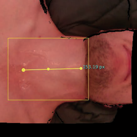
First results of a clinical study (see publication below) demonstrated that robotic ultrasound-assisted facet joint insertions lead to success rates comparable to current clinical practice while lowering the X-ray dose and offering additional anatomical context for needle trajectory planning.
Some videos:
Related Publications
2018
Robotic ultrasound-guided facet joint insertion
Javier Esteban, Walter Simson, Sebastian Requena Witzig, Anna Rienmüller, Salvatore Virga, Benjamin Frisch, Oliver Zettinig, Drazen Sakara, Yu-Mi Ryang, Nassir Navab, and Christoph Hennersperger
International Journal of Computer Assisted Radiology and Surgery, Jun 2018
Purpose: Facet joint insertion is a common treatment of chronic pain in the back and spine. This procedure is often performed under fluoroscopic guidance, where the staff’s repetitive radiation exposure remains an unsolved problem. Robotic ultrasound (rUS) has the potential to reduce or even eliminate the use of radiation by using ultrasound with a robotic-guided needle insertion. This work presents first clinical data of rUS-based needle insertions extending previous work of our group. Methods: Our system implements an automatic US acquisition protocol combined with a calibrated needle targeting system. This approach assists the physician by positioning the needle holder on a trajectory selected in a 3D US volume of the spine. Results: By the time of submission, nine facets were treated with our approach as first data from an ongoing clinical study. The insertion success rate was shown to be comparable to current clinical practice. Furthermore, US imaging offers additional anatomical context for needle trajectory planning. Conclusion: This work shows first clinical data for robotic ultrasound-assisted facet joint insertion as a promising solution that can easily be incorporated into the clinical workflow. Presented results show the clinical value of such a system.
2017
Towards MRI-Based Autonomous Robotic US Acquisitions: A First Feasibility Study
Christoph Hennersperger, Bernhard Fuerst, Salvatore Virga, Oliver Zettinig, Benjamin Frisch, Thomas Neff, and Nassir Navab
On the reproducibility of expert-operated and robotic ultrasound acquisitions
Risto Kojcev, Ashkan Khakzar, Bernhard Fuerst, Oliver Zettinig, Carole Fahkry, Robert DeJong, Jeremy Richmon, Russell Taylor, Edoardo Sinibaldi, and Nassir Navab
International Journal of Computer Assisted Radiology and Surgery, Jun 2017
3D ultrasound registration-based visual servoing for neurosurgical navigation
Oliver Zettinig, Benjamin Frisch, Salvatore Virga, Marco Esposito, Anna Rienmüller, Bernhard Meyer, Christoph Hennersperger, Yu-Mi Ryang, and Nassir Navab
International Journal of Computer Assisted Radiology and Surgery, Sep 2017
Purpose: Precise needle placement is an important task during several medical procedures. Ultrasound imaging is often used to guide the needle toward the target region in soft tissue. This task remains challenging due to the user’s dependence on image quality, limited field of view, moving target, and moving needle. In this paper, we present a novel dual-robot framework for robotic needle insertions under robotic ultrasound guidance. Method: We integrated force-controlled ultrasound image acquisition, registration of preoperative and intraoperative images, vision-based robot control, and target localization, in combination with a novel needle tracking algorithm. The framework allows robotic needle insertion to target a preoperatively defined region of interest while enabling real-time visualization and adaptive trajectory planning to provide safe and quick interactions. We assessed the framework by considering both static and moving targets embedded in water and tissue-mimicking gelatin. Results: The presented dual-robot tracking algorithms allow for accurate needle placement, namely to target the region of interest with an error around 1 mm. Conclusion: To the best of our knowledge, we show the first use of two independent robots, one for imaging, the other for needle insertion, that are simultaneously controlled using image processing algorithms. Experimental results show the feasibility and demonstrate the accuracy and robustness of the process.
Automatic force-compliant robotic ultrasound screening of abdominal aortic aneurysms
Salvatore Virga, Oliver Zettinig, Marco Esposito, Karin Pfister, Benjamin Frisch, Thomas Neff, Nassir Navab, and Christoph Hennersperger
In 2016 IEEE/RSJ International Conference on Intelligent Robots and Systems (IROS), Oct 2016
Ultrasound (US) imaging is commonly employed for the diagnosis and staging of abdominal aortic aneurysms (AAA), mainly due to its non-invasiveness and high availability. High inter-operator variability and a lack of repeatability of current US image acquisition impair the implementation of extensive screening programs for affected patient populations. However, this opens the way to a possible automation of the procedure, and recent works have exploited the use of robotic platforms for US applications, both in diagnostic and interventional scenarios. In this work, we propose a system for autonomous robotic US acquisitions aimed at the quantitative assessment of patients’ vessel diameter for abdominal aortic aneurysm screening. Using a probabilistic measure of the US quality, we introduce an automatic estimation of the optimal pressure to be applied during the acquisition, and an online optimization of the out-of-plane rotation of the US probe to maximize the visibility of the aorta. We evaluate our method on healthy volunteers and compare the results to manual acquisitions performed by a clinical expert, demonstrating the feasibility of the presented system for AAA screening.
Toward real-time 3D ultrasound registration-based visual servoing for interventional navigation
Oliver Zettinig, Bernhard Fuerst, Risto Kojcev, Marco Esposito, Mehrdad Salehi, Wolfgang Wein, Julia Rackerseder, Edoardo Sinibaldi, Benjamin Frisch, and Nassir Navab
In 2016 IEEE International Conference on Robotics and Automation (ICRA), May 2016


 On the reproducibility of expert-operated and robotic ultrasound acquisitionsInternational Journal of Computer Assisted Radiology and Surgery, Jun 2017


